
I have been working in Neuroscience field for almost 10 years. In this field, early diagnostics of neurodegeneration was always a dream for many researches. Existing biophysical approaches offer many advantages, but they also have serious limitations, such as spatial or temporal resolution, invasiveness, toxicity, poor specificity, etc. Hyperspectral imaging of retina raises hopes that we finally can revolutionize diagnostics of brain neurodegeneration.
Early diagnostics of neurodegeneration is a key to improve lives of millions of people. Have we finally found the way to do it? A Canadian company, Optina Diagnostics is developing a tool to study neurodegeneration by using hyperspectral imaging of retina. It is a viable approach because retina shares a lot of functional similarities with the brain, however, the analysis of retinal hyperspectral images is technically challenging. That problem can be solved using Artificial Intelligence (AI). The combination of hyperspectral imaging and AI-based analysis algorithm recently started being tested for clinical diagnostic of neurodegeneration (Alzheimer’s disease). In this post I am investigating whether this combination has a potential to be efficient in patients to diagnose neurodegeneration.
Neurodegenerative disorders increasingly become an issue in many developed countries around the world and cause 12% of deaths in Europe. In France, the total number of people suffering from different neurodegenerations (Alzhneimer’s, Parkinson’s, Huntington’s diseases, Epilepsy, Multiple sclerosis, Brain tumors, etc.) ads up to 1.6 million and this number is constantly growing. However, reliable treatments or timely diagnostics for neurodegenerative disorders are not well developed yet. Often, when symptoms become evident it is too late to prevent the neurodegeneration. Therefore, researchers are looking for noninvasive early diagnostics strategies.
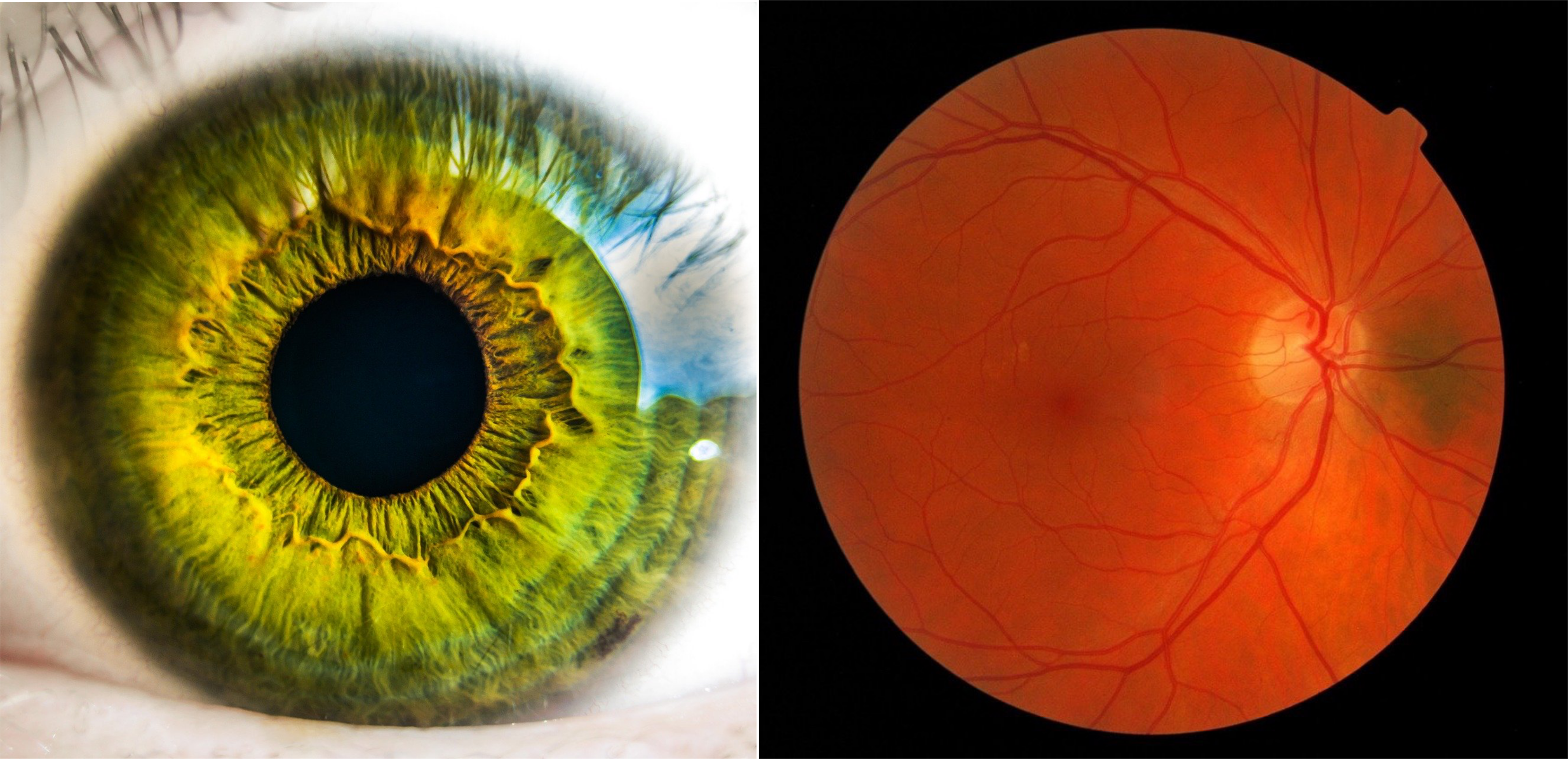
An optical Imaging of retina can be one of the ways for efficient and noninvasive brain diagnostics. Retina is easily accessible and has a unique feature sharing the same origin as central nervous system. Since retina anatomically and developmentally is connected to the brain, retinal imaging can reveal to us accumulation of certain molecules in the brain (molecular biomarkers of neurodegeneration)[1]. Currently, this approach proofed itself very useful in diagnostics of retinal lesions, change in oxygen supply[2], retinopathy, retinal vein occlusions (RVOs), glaucoma, etc.[3] On the side of improving data analysis, the current trend of using AI and Machine Learning (ML) for biological problems also showed impressive results increasing accuracy and assisting medical doctors identify bone fractures[4], cancer [5], rare diseases[6] etc. Currently, many retinopathic conditions were analysed with good accuracy using ML. Some published algorithms were very successful in detection of visible by human eye pathologies (hemorrhages, drusen, exudates, etc.). Using ML for more subtitle conditions as early accumulation of amyloid beta, etc. is a less trivial task. Optina Diagnostics (http://optinadx.com/) takes this challenge and develops the combination of hyperspectral imaging and AI-based analysis to provide the necessary solution for early diagnostic of neurodegeneration.
Retinal imaging (hyperspectral) is now used by Canadian company Optina Diagnostics to develop retinal phenotyping and Alzheimer’s disease diagnostic as the first step. Hyperspectral imaging measures spectral characteristics (absorption/reflection of light) of retina at different wavelengths in 450-900 nm range by. This spectrum can change in pathological conditions Hyperspectral imaging has a number of advantages including lower cost compare to positron emission tomography (PET), it does not require additional markers, it is relatively noninvasive and it allows identifying specific molecules in certain conditions. The output data of hyperspectral imaging are represented as spectral characteristics of individual pixels in a set of frames for each wavelength (so called - hypercube), that in total, gives a large volume of data that is hard to handle. In this paper[7] the approach is to reduce to “reflectance spectra” and add Dimension Reduction by Orthogonal Projection algorithm to improve identification of amyloid beta. This method showed a statistical difference under amyloid beta accumulation[8] with classic analysis approach. Optina Diagnostics wants to use the combination with custom-made algorithm Artificial Intelligence-based (AI) analysis might become useful to predict early onset of Alzheimer’s and classify other diseases from single set of images from one patient. Optina Diagnostics developed an AI-based classifier predicting the presence of amyloid beta with up to 90% accuracy. However, high accuracy does not always say that efficiency will be as high. Thus, the question remains whether the combination of hyperspectral imaging and AI-based analysis going to be efficient in patients because there are several serious challenges lie on the way.
While hyperspectral imaging of retina and AI-based analysis offers hopes for easier and faster early diagnostics of neurodegeneration, there are several challenges on this way. First, this approach might not be sensitive enough to distinguish the state of amyloid beta molecules (oligomers, polymers, aggregates). While it is crucial for early diagnostics to catch oligomers which appear to be one of the most critical steps triggering neurodegenerations. The utilization of specific fluorescent markers could serve as a good alternative way of diagnostics for neurodegenerative diseases. However, currently we are lacking high quality markers. Second, the cost of the camera for hyperspectral imaging is very high for broad market and data analysis process is very complex while the results are highly variable depending on the specific location of retina. This solution might not be so universal and could still require additional testing with traditional methods (PET, cerebral spinal fluid tests, etc.). Third, amyloid beta as a biomarker for Alzheimer’s disease it can also be a sign of other conditions, e.g. cancer. This means, that identification of its accumulation does not necessarily say which disease it is and which treatment has to be used. Forth, the data obtained with hyperspectral imaging reflects a complex signal contributed from hundreds of molecules. Specific biomarkers for different diseases could represent only very minor part of overall signal and therefore could be distinguished only by using sophisticated algorithms. The interference between different contributing molecules is also hardly distinguishable. AI-based algorithms often functioning as a “black box” might not be the most suitable solution to solve this complex task.
Overall, at current stage, the choice of hyperspectral imaging + AI might not be the most optimal for precise identification of amyloid beta accumulation or other biomarkers for brain pathologies. For the moment, this method is more efficient for identification of visible pathologies related to vascularization, rather than subtitle accumulation of certain molecules. The good news is that with further development of this technology and AI algorithms we might be one step closer to early diagnostic of neurodegeneration and an improvement of many lives around the globe.
Please, if you like this article, please share it with your friends, subscribe to our news and free networking events here and leave your comments below.
[1.] Chiquita, S. et al. The Retina as a Window or Mirror of the Brain Changes Detected in Alzheimer’s Disease: Critical Aspects to Unravel. Mol. Neurobiol. 56, 5416–5435 (2019).
[2.] Mordant, D. J. et al. Spectral imaging of the retina. Eye 25, 309–320 (2011).
[3.] Reshef, E. R., Miller, J. B. & Vavvas, D. G. Hyperspectral Imaging of the Retina: A Review. Int. Ophthalmol. Clin. 60, 85–96 (2020).
[4.] Lindsey, R. et al. Deep neural network improves fracture detection by clinicians. Proc. Natl. Acad. Sci. U. S. A. 115, 11591–11596 (2018).
[5.] Azuaje, F. Artificial intelligence for precision oncology: beyond patient stratification. npj Precis. Oncol. 3, 1–5 (2019).
[6.] Stafford, I. S. et al. A systematic review of the applications of artificial intelligence and machine learning in autoimmune diseases. npj Digit. Med. 3, (2020).
[7.] Hadoux, X. et al. Non-invasive in vivo hyperspectral imaging of the retina for potential biomarker use in Alzheimer’s disease. Nat. Commun. 10, 1–12 (2019).
[8.] Koronyo, Y. et al. Retinal amyloid pathology and proof-of-concept imaging trial in Alzheimer’s disease. JCI insight 2, 1–19 (2017).