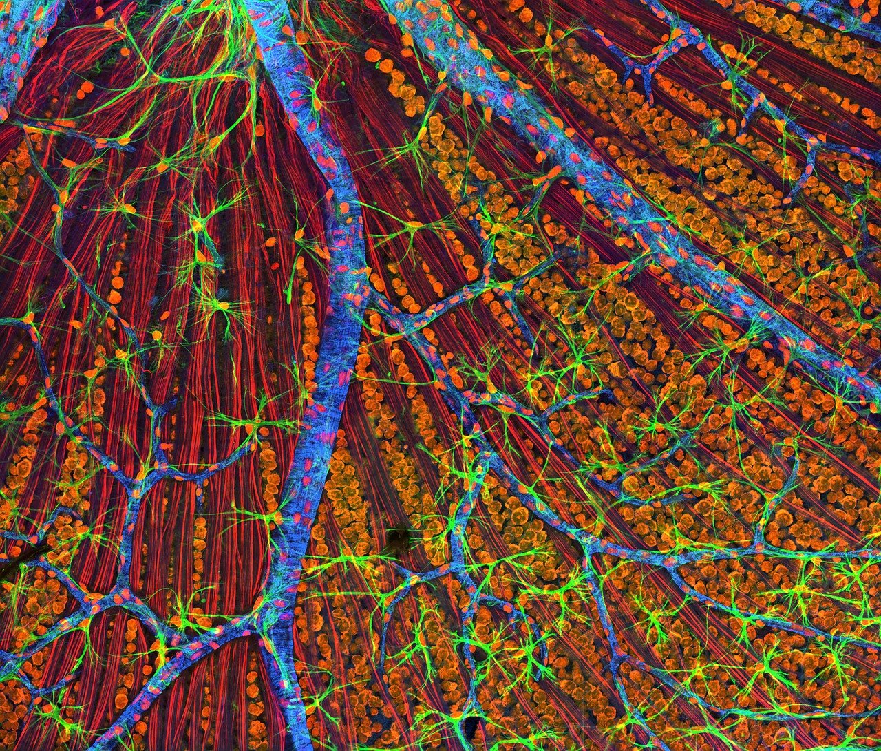


Optical Coherence Tomography (OCT) is a biomedical optical imaging technique that uses low coherence interferometry to resolve backscattered light by depth. It has become a widely used technique for ophthalmology for its unique ability to non-invasively assess the various layers of the retina. However, the high cost of clinical OCT systems (up to $150,000) has limited access to mostly large eye centers and laboratories. We have chosen to pursue a low-cost OCT system to increase patient access, particularly in low cost settings. To further increase access we have developed a highly portable and robust system, capable of operating at the point of care. This talk will present design and applications of low-cost OCT, including identifying retinal biomarkers of Alzheimer’s disease.
Author Dr. Adam Wax, Dept. of Biomedical Engineering, Duke University. Here is more information.
OCT is an optical analogue of Ultrasound imaging. You send light into the tissue and measure the back-scattered reflections. By noting the time of flight or how deep the light has penetrated you can map out the three-dimensional tissue profile. For example, in clinical practice, OCT was of a particular use in ophthalmology. OCT can help to identify changes in thickness of retina and diagnose retinal degradation that causes vision loss. However, existing OCT systems are quite expensive (+35K$) and might not be always available in less developed countries.
We have developed our first OCT system couple of years ago for the price ~7K$ (here is more info ). We have borrowed high-performance, but low-cost optical parts from other applications and optimized them for our system to reduce the price without significant loss of quality. We brought down the price further by using 3D printed parts. It also gave our system flexibility that is not available for other systems. We can easily adjust our system for different applications. Our system has also relatively small size and light weight (~3 kg) compared to other similar systems. Most importantly, the performance matched the level of other entry-level OCT systems. The quality of obtained images was comparable to those obtained with state-of-the-art OCT systems.
Our second-generation system cost ~5K$ and the weight ~1.8kg with integrated PC (here is more info ). The performance was similar to the OCT system from Heidelberg engineering. The images from our system were slightly dimmer, but al layers were visible and sufficiently resolved for diagnostic. While the parameters of systems were similar, our system was significantly smaller, lighter and way more attractive in terms of the price (5K$ vs. 60K$). Regarding the performance the Heidelberg engineering’s system had 6% better signal-to-noise ratio. However, considering the price reduction, these 6% difference is a reasonable trade-off.
At Lumedica we are offering OQLabScope2.0 for <10k$ (link) and we have distributors in Europe. We are working now on our clinical system OQ Eyescope1.0 that is available for research purposes and soon will be approved for clinical use on humans. Our systems are very agile and can be relatively easy modified for different uses. Our modular design allows rapidly change imaging parameters (e.g. resolution, canter wavelength and wavelength range) by changing the grating, sensor and lens tube.
Standard OCT systems are not suitable during a surgery. We adapted an OCT system for surgical access. We placed an OCT into a commercial rigid borescope, a tool that is used for minimal invasive surgeries e.g. laparoscopy. We attached it to our OCT systems by using custom 3D printed parts. In his combination our OCT resolution (axial- 8.2 microns, lateral – 10.3 microns) was similar to those of other available systems. We studied the thickness of articular cartilage on Porcine Femur bone and measured it with a very high precision. This is particularly useful during the surgery when standard methods such as fMRI could not be applied anymore. Having a small probe during the surgery allows the online monitoring of 3D tissue profile. With a simple false-colour mapping it was possible to mark the areas with different thickness of cartilage and help the doctor to navigate during the surgery.
Another area that we are working on is gastro-intestinal applications, e.g. esophagus. Is the part of gastro-intestinal system where in the recent years the occurrence of cancer significantly increased. We significantly modified parameters of our OCT system and attached it to a rotary probe that inserts into esophagus and rotates by a motor around and images the esophagus around in a circle. Some parts of the probes were 3D printed. Optical parts of the probe were assembled and consisted of a single-mode fibre, a GRIN lens and a prism that turns the light beam. These parts were placed into a 3D printed scanhead adapted for endoscopic applications. Thanks to our 3D printing technique we were able to easily modify the parameters of the scanhead saving weeks for adjustments. We created 2 generations of probes and improved the image quality and reduced reflections. In phase I of clinical trials (finished 02/2020) we looked at Barrett’s esophagus and we could clearly see the typic changes in tissue. Phase 2 clinical trials are ongoing to identify signs of tissue alteration in precancer patients.
Cervical cancer is the most common type of gynaecological cancer. The most abundant diagnostic tool for it is pap smear. The limitation of this method is time consumption, resource cost and limited specificity where there are many false positives occur. Then it requires biopsy before the diagnose can be validated. Our approach is to use angle-resolved low-coherence interferometry (a/LCI). Me measure scattered light as a function of scattering angle and this lets us determine the size of the scatters. Here are the cell nuclei and by measuring their enlargement we can identify precancerous lesions with high specificity and good sensitivity. We did a clinical study and showed that we could detect precancer or dysplasia in vivo. Now we are developing a probe for cervical cancer detection. Since nuclear diameter can be a marker for dysplastic changes, measuring enlargement of nuclei from 8-10 microns to 12-15 microns can predict precancerous lesions development. Usually OCT cannot resolve nuclei on its own so we have to use angular scattering. Confocal microscopy could also be used for the task, but it requires contrasting agents and limited in depth. a/LCI enables identification of changes in nuclei size in deep layers (300-400 microns below the surface) which are the most valuable for diagnostic. We have developed a specific probe for a/LCI and measured the nuclei size with high precision (0.25 microns error) and have finished clinical study with it. The sensitivity of this method is 100%, meaning that a doctor can be sure that there is no cancer in the sample.
Alzheimer’s disease is a common neurodegenerative disease that is characterized by accumulation of amyloid beta plaques. Symptoms of Alzheimer’s disease include memory loss and cognitive decline. Currently there is no reliable way to detect Alzheimer’s disease. Often it is done by cognitive tests that are very subjective. It can be diagnosed with combination of fMRI and CT, but it is costly (2-5K$ per patient). Early and reliable detection of Alzheimer’s disease is in high demand because sooner the patient receives the treatment, better results can be expected. OCT can provide efficient, non-invasive early diagnostic tool for Alzheimer’s disease. Measuring the properties of retinal tissue offers us a solution, because retina is also a neural tissue, but it exposed to optical interrogation. We combined OCT with a/LCI to study retina. Taking simultaneously both measurements from retina allowed us to identify the changes in retinal layers thickness in transgenic mice (Alzheimer’s disease animal model) as it was previously shown. Most importantly, we have identified the changes in light scattering patterns in Alzheimer’s mice compared to wild type mice suggesting a rougher surface of retina. These properties can offer a solution for early diagnostic of Alzheimer’s disease. Currently we are working on validation of these results in humans.
Low-cost OCT is comparable with its properties to other commercially available systems. Low-cost OCT was validated in clinical trials Our low-cost OCT is an adjustable system that can be easily modified for different applications (Gynaecology, Orthopaedics and Neurology).
Please, if you like this article, please share it with your friends, subscribe to our news and free networking events here and leave your comments below.
[1.] The talk of Dr. Adam Wax: Low-cost, Portable Optical Coherence Tomography for Point of Care Use.
[2.] Design and implementation of a low-cost, portable OCT system. Sanghoon Kim, Michael Crose, Will J. Eldridge, Brian Cox, William J. Brown, and Adam Wax. Biomedical Optics Express. Vol. 9, Issue 3, pp. 1232-1243 (2018) (link)
[3.] Lumedica creates affordable light-based scientific and medical instruments that deliver accurate diagnostic results. Lumedica
[4.] First clinical application of low cost portable OCT system. Ge Song, Sanghoon Kim, Michael Crose, Brian Cox, Evan Jelly, J. Niklas Ulrich, Adam Wax. Proceedings Volume 10858, Ophthalmic Technologies XXIX.(2019) (link)
[5.] The OQ LabScope enables researchers, innovators and educators to affordably access OCT’s powerful imaging capability.(link)